
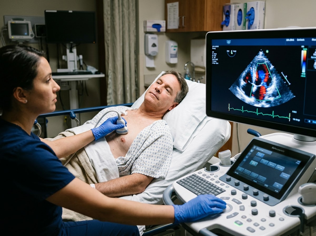
What is a 2D Echo Test?
A 2D Echo test — full name two-dimensional echocardiography, also called an echocardiogram or TTE (transthoracic echocardiography) — is a heart ultrasound. It works the same way as the ultrasound used to see a baby during pregnancy. A small probe placed on your chest sends sound waves into the heart, and the echoes that bounce back form a live, moving picture on the screen.
The cardiologist can watch all four heart chambers, the four heart valves opening and closing, the thickness of the heart walls and how strongly the heart pumps. A Doppler study is done in the same sitting. Doppler shows the direction and speed of blood flow, so the doctor can see a leaking valve, a narrowed valve or high pressure inside the heart.
Think of it like this: if the heart is a house, an ECG checks the electrical wiring. A 2D Echo looks at the walls, rooms and doors — how big the rooms are, how thick the walls are and whether the doors (valves) open and shut properly.
Why is a 2D Echo done?
A 2D Echo is done to see the structure and heart function directly — something an ECG or a blood test cannot do. One of the most useful numbers in the report is the ejection fraction (EF). In simple words, EF tells how much of the blood in the main pumping chamber is pushed out with each beat. In most adults an EF of around 55% or more is considered normal; a lower EF means the heart is pumping weakly. That is why the 2D Echo is often called the heart pumping test.
When is an Echo Test Needed?
Doctors ask for a 2D Echo when a symptom or a finding could come from the heart muscle or valves, and to keep track of a known heart problem over time.
Symptoms where a 2D Echo test is useful
Who needs a 2D Echo?
- People with breathlessness or a suspected weak heart (echo for weak heart)
- Anyone with a murmur or a known heart valve problem
- After a heart attack, to check how much the muscle is affected
- Long-standing high BP or diabetes, to look for heart strain
Other common reasons
- Before major surgery, to check heart fitness
- Follow-up of heart failure or valve disease treatment
- Changed ECG or an enlarged heart on chest X-ray
- Pregnancy with heart symptoms or past heart disease
Severe breathlessness or chest pain right now? Do not wait for a scheduled Echo. Sudden breathlessness, chest pain with sweating, or fainting needs emergency care first. Call +91 77778 65621 or come to our 24x7 cardiac emergency.
What Does 2D Echo Detect?
The 2D Echo is a complete cardiac imaging check of the heart’s structure and function. These are the main problems it can show:
Weak heart (low EF)
A low ejection fraction shows the heart is not pumping well, as in heart failure or cardiomyopathy.
Heart failure careHeart valve problems
Narrowed (stenosis) or leaking (regurgitation) valves, including rheumatic valve disease — Doppler measures how severe it is.
Heart valve diseaseDamage after a heart attack
Parts of the heart wall that move less than normal point to muscle that has lost blood supply.
Heart attack treatmentHoles and birth defects
Holes in the heart (ASD, VSD) and other congenital problems can be seen in adults and children.
Congenital heart diseaseThick or enlarged heart
Thickened walls from long-term high BP, or stretched, enlarged chambers.
Fluid, clots and lung pressure
Fluid around the heart (pericardial effusion), some blood clots inside the chambers, and raised pressure in the lung arteries.
What a 2D Echo cannot tell: a resting Echo does not show the heart arteries themselves, so it cannot rule out a blockage. If your chest pain comes on exertion, your doctor may add a TMT or Stress Echo. If the pictures are not clear enough, or a detailed view of a valve or clot is needed, a TEE may be advised. For babies and children, see pediatric echo.
How is a 2D Echo Test Done?
How long does a 2D Echo take? Usually about 20 to 30 minutes, sometimes a little longer if more Doppler measurements are needed. You stay awake and can go home or back to work straight after.
You change and lie on your left side
You are given a gown that opens at the front and asked to lie on a couch, turned towards your left side. This brings the heart closer to the chest wall for clearer pictures. Women patients can ask for a female staff member to be present.
ECG stickers are placed
Three small stickers on the chest connect to the Echo machine, so each picture can be matched to the timing of your heartbeat.
Gel and probe on the chest
A little cool, water-based gel is put on the chest and the probe is gently moved over different spots — near the breastbone, below the left breast and under the ribs.
Doppler and measurements
You may be asked to hold your breath for a few seconds or turn slightly. During Doppler you may hear a whooshing sound — that is simply your blood flow, and it is normal.
Gel wiped off, report prepared
The gel is wiped away and you can dress. The cardiologist reviews the images and measurements, and your doctor will tell you when the report and explanation will be ready.
No radiation, no current, no injection. Echo uses sound waves, not X-rays. It is safe for pregnant women, children and elderly patients, and can be repeated as often as your doctor needs.
Preparation for a 2D Echo — and is it Safe?
A standard 2D Echo needs no special preparation. These small things simply make the test smoother.
Good to do
- Wear a two-piece outfit so only the top needs to be removed
- Bring old Echo, ECG and discharge reports, and your medicine list
- Eat normally and take your regular medicines
- Tell the staff if you cannot lie on your left side because of pain
Better to avoid on the test day
- Stopping BP or heart medicines on your own
- Oil, lotion or powder on the chest
- Heavy chains or necklaces
- Talking or moving a lot while images are taken
Is 2D Echo painful?
No. The gel feels cool and you will feel light pressure from the probe. In a few spots, such as under the ribs, the doctor may press a bit firmer to get a clear view — tell them if it feels uncomfortable. There are no needles and no radiation, and there are no known side effects of a standard 2D Echo.
2D Echo vs ECG — What is the Difference?
Patients often get both tests on the same day and wonder why. They look at two different sides of the heart and work best together.
| 2D Echo | ECG | |
|---|---|---|
| What it checks | Structure and pumping: chambers, valves, walls, EF | Electrical activity: rate and rhythm |
| How it works | Ultrasound probe with gel on the chest | Stickers on chest, arms and legs |
| Time taken | About 20–30 minutes | About 5–10 minutes |
| Pain or radiation | None | None |
| Best for | Breathlessness, weak heart, murmur, valve problems | Chest pain, palpitations, heart attack signs |
| What it can miss | Blockage in heart arteries | Valve problems and pumping strength |
In short: the ECG tells how the heart is beating; the 2D Echo tells how the heart is built and pumping. Your cardiologist decides which one you need, or both.
What Happens After Your 2D Echo Report?
Your report lists the size of each chamber, wall thickness, how each valve is working, Doppler findings and the EF. Our cardiologist reads it together with your symptoms, ECG and other reports and explains it in plain words. Usually one of three things follows:
- Normal Echo: the heart’s structure and pumping are fine. You get advice on BP, sugar, cholesterol and lifestyle, and further tests only if your symptoms need them.
- Mild changes: such as a mild valve leak or slight wall thickening. These are often watched with medicines and a repeat Echo after some time, as your doctor advises.
- Significant problem: such as a low EF or a tight valve. Your doctor will plan treatment — medicines, further tests or a procedure — and explain each option.
Tip: keep your Echo reports in one file and bring them to every visit. Comparing today’s EF and valve findings with older reports shows whether things are improving or need a change in treatment.
Why Get Your 2D Echo Test at Prathmesh Cardiac Institute?
An Echo is only as useful as the person reading it. Here, your images are reviewed by the same heart team that treats heart failure, valve disease and heart attacks.
Reviewed by a DM cardiologist
Your Echo is interpreted by a heart specialist who looks at it alongside your symptoms and history.
Explained in simple words
You are told what your EF, valves and chambers show, and what it means for you.
Doppler in the same sitting
Blood flow, valve leaks and pressures are checked during the same test, with no second visit.
Next test under one roof
ECG, TMT, Stress Echo, TEE and Holter are available here if your doctor needs more information.
24x7 cardiac backup
If the Echo shows something urgent, our CCU and cath lab are in the same building.
Easy to reach in Raipur
NH-30, Amlidih Road, Deopuri — convenient for patients from Raipur and nearby districts.
Who Reads Your Report
Your report is studied by a DM Cardiology specialist, who explains it to you in plain words and tells you what to do next.
800 × 1000 px · JPG

Dr. Gaurav Tripathi
Treats heart attacks and blocked arteries, and uses Echo findings to plan angioplasty and follow-up care.
800 × 1000 px · JPG

Dr. Sulabh Chandrakar
Sees patients with breathlessness, valve problems and heart failure, and explains Echo reports in detail.
2D Echo Test — Questions Patients Ask Us
A 2D Echo test is an ultrasound of the heart. A probe with gel is moved over your chest and sound waves create a live picture of the heart chambers, valves and walls, showing how well the heart is pumping.
It is done to check the structure and pumping strength of the heart. It helps find the cause of breathlessness, chest discomfort, swelling of the feet or a heart murmur, and to follow up known heart failure or valve disease.
A 2D Echo can detect a weak heart (low EF), narrowed or leaking valves, damage after a heart attack, holes in the heart, a thickened or enlarged heart, fluid around the heart, some clots inside the heart and raised pressure in the lungs.
An Echo test is needed when you have breathlessness, chest discomfort, swelling of the feet, fainting or a murmur, after a heart attack, or before major surgery. It is also repeated to track a known heart problem, as your doctor advises.
No. You only feel cool gel and light pressure from the probe. There are no needles, no injection and no radiation, and you can go back to normal work right after the test.
A 2D Echo usually takes about 20 to 30 minutes. It can take a little longer if detailed Doppler measurements of the valves are needed.
An Echo shows the size of the four heart chambers, how the four valves open and close, how thick the heart walls are and how strongly the heart pumps (ejection fraction). Doppler adds the direction and speed of blood flow through the heart.
People with breathlessness, a suspected weak heart, a heart murmur, valve disease, a recent heart attack, long-standing high BP or abnormal ECG findings usually need a 2D Echo. Your cardiologist will tell you if and when you need one.
Ejection fraction is the share of blood the main pumping chamber pushes out with each beat. In most adults about 55% or more is considered normal; a lower value means the heart is pumping weakly. Your doctor will explain what your own number means.
No. You can eat, drink and take your regular medicines before a standard 2D Echo. Fasting is needed only for a TEE, which is a different test.