
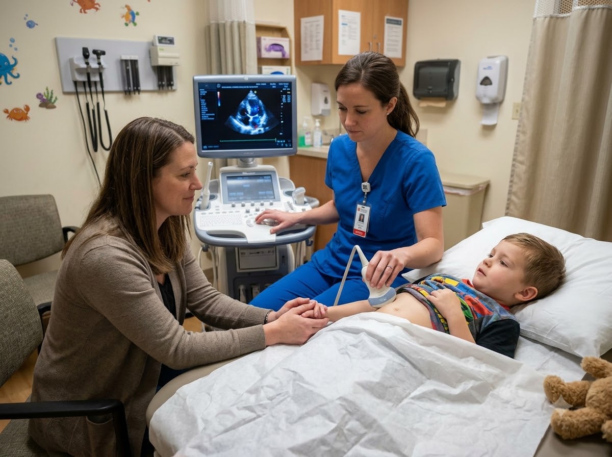
What is a Pediatric Echo?
A pediatric echo (pediatric echocardiography, or an echocardiogram for children) is an ultrasound scan of a child’s heart. A small probe with cool gel is moved over the chest. Sound waves bounce off the heart and make a live moving picture on the screen — the same kind of ultrasound used to see a baby during pregnancy.
The doctor can see the heart structure clearly: the four heart chambers, the heart valves, the walls between the chambers and the big blood vessels. Colour Doppler shows the direction of blood flow, so a small hole or a leaking valve stands out. The same test, done for grown-ups, is the routine 2D Echo — and we do both here, for adults and for children.
Think of it like this: an ECG checks the heart’s electrical wiring. An echo looks at the building itself — the rooms, the doors (valves), the walls between the rooms, and whether the pump is working well.
Why is pediatric Echo done?
Most heart problems in children are things they are born with — a congenital heart defect such as a hole in the heart or a narrow valve. A child cannot describe these problems, and many show only small signs like a murmur, sweating during feeds or slow weight gain. A pediatric echo answers the main question for the family: is the heart normal, or does it need care? In most children with an innocent murmur, the echo simply gives peace of mind.
When Does a Child Need an Echo?
Usually the child’s paediatrician hears something during a routine check, or the parents notice a change in feeding, breathing or growth. These are the common reasons a child heart test is advised.
Signs in a baby or child where an echo helps
Heart test for newborn and baby
- A murmur heard after birth or at a vaccination visit
- Low oxygen reading or bluish colour after birth
- A doubt raised in the pregnancy scan (fetal heart)
- Babies with Down syndrome or other genetic conditions, as advised by the paediatrician
Heart checkup for kids and adults
- Older child with breathlessness, tiredness or chest discomfort on play
- Repeated chest infections with slow growth
- Joint pains and fever in the past (to check for rheumatic heart disease)
- Adults: breathlessness, chest pain, swelling of feet, high BP or a valve problem
Go to an emergency now if a baby turns blue, breathes very fast and struggles, stops feeding, becomes very drowsy or a child faints. Call +91 77778 65621 — our cardiac emergency is open 24x7.
What Does a Pediatric Echo Detect?
The echo shows both the structure of the heart and how well it pumps. These are the common findings in children — and the same scan in an adult checks the valves and pumping strength.
Hole in the heart (ASD, VSD)
A gap in the wall between the upper (ASD) or lower (VSD) chambers. The echo shows its size and whether it may close on its own.
Congenital heart diseaseOpen blood vessel (PDA)
A small vessel that should close soon after birth but stays open. Common in premature babies.
Learn about PDANarrow or leaking valves
Valves born narrow, or valves damaged by rheumatic fever in older children and adults.
Heart valve diseaseComplex defects (“blue baby”)
Conditions such as Tetralogy of Fallot, where mixing of blood causes blue lips or nails. The echo gives the first clear picture.
Weak heart muscle
Poor pumping (low EF) from cardiomyopathy or heart muscle inflammation, in children and in adults.
Heart failure careFluid and pressure changes
Fluid around the heart, or raised pressure in the lung blood vessels, which can follow a long-standing hole.
What an echo does not show: it does not record the heart rhythm over time — for a child with a fast heartbeat that comes and goes, the doctor may add an ECG or Holter monitoring. For some complex defects, further scans such as a CT or MRI may be advised.
How is an Echo Done in Children and Adults?
The scan usually takes 20 to 40 minutes. For a small child we take a little extra time so the child is calm — a quiet, settled child gives the clearest pictures.
Short talk with the doctor
We ask about birth, feeding, growth, breathing and any earlier reports. Bring the baby’s discharge papers and pregnancy scan reports if you have them.
Child lies down, parent stays close
Small babies can lie on the bed or on a parent’s lap. A parent stays in the room through the whole test.
Gel and a small probe on the chest
Some gel is applied and a small probe is moved over the chest and below the ribs. Nothing goes inside the body.
Keeping the child calm
A feed, a favourite toy or a cartoon on the phone helps. If a very small child cannot settle, the doctor may suggest a mild sedative given under supervision.
Doctor explains the findings
The gel is wiped off and the cardiologist explains what was seen in simple words, and what the next step is.
For adults the steps are the same: you lie on your left side, gel is applied and the probe is moved over the chest. It is the standard 2D Echo — painless and with no radiation.
Preparing Your Child for an Echo
No blood test, no injection and usually no fasting is needed. A little planning helps a child stay calm.
Good to do
- Dress the child in loose, front-opening clothes
- Bring a toy, a blanket or a bottle; for infants, time the feed close to the test
- Bring all old reports, birth papers and the pregnancy scan
- Tell older children simply: “It is like a scan with cold jelly, it will not hurt”
Better to avoid
- Arriving in a rush — a tired, hungry child is harder to settle
- Body oil or powder on the chest
- Scaring the child with words like “injection”
- Giving any sleep medicine at home on your own
Is Echo Safe for Children?
Yes. Echo uses sound waves, not X-rays, so there is no radiation. It is safe for newborns and premature babies and can be repeated as often as needed. There are no needles. The only discomfort is the cool gel and some pressure from the probe. If a sedative is ever needed for a very small child, it is given only when the doctor feels it is necessary, with the child watched until fully awake, and you will be told in advance about feeding times.
Echo for Children vs Echo for Adults
It is the same machine and the same basic method. What changes is how the test is planned and what the doctor is looking for.
| Child or baby | Adult | |
|---|---|---|
| Preparation | Feeding and sleep timed so the child is calm; toy or cartoon helps | Usually none |
| Sedation | Sometimes, for a very small child who cannot settle — only if the doctor advises | Not needed |
| Who is in the room | A parent stays with the child throughout | Patient alone, or with an attendant if wished |
| Probe | A smaller probe suited to a small chest | A standard adult probe |
| Main things looked for | Holes in the heart, open vessels, narrow valves, how the heart is formed | Pumping strength (EF), valve narrowing or leak, damage after a heart attack |
| Time | May take longer if the child needs time to settle | Usually 15–30 minutes |
For parents: crying makes the pictures less clear, not the test less safe. If your child is upset, we pause, let you comfort them, and continue.
What Happens After the Echo Report?
The cardiologist looks at the echo together with your child’s growth, symptoms and examination. Usually one of three things follows:
- Normal heart, innocent murmur: many murmurs in children are harmless sounds of normal blood flow. No treatment and no restrictions are needed.
- A small defect that may close by itself: many small holes close as the child grows. The doctor plans a follow-up echo after some months.
- A significant or complex defect: the family is guided clearly on the next steps — medicines, timing, and whether a catheter procedure or surgery is needed, and where it can best be done.
For adults, the report is used in the same way to plan treatment for valve problems, a weak heart or blocked arteries. Read more about congenital heart disease and its treatment.
Tip: keep every echo report of your child in one file. Comparing today’s echo with an older one shows whether a hole is getting smaller or a valve is changing.
Why Choose Prathmesh Cardiac Institute for an Echo?
A heart test for a child worries the whole family. We keep it calm, unhurried and clearly explained.
Done and read by DM cardiologists
The echo is performed and interpreted by heart specialists with DM Cardiology training.
Parents stay with the child
You remain in the room throughout, and the test is paused whenever the child needs comfort.
Unhurried, child-friendly approach
We give small children time to settle, because a calm child gives the clearest pictures.
Clear guidance if a defect is found
You are told what the finding means, whether it can wait, and what the next step should be.
Adult echo and other tests here
Echo for adults, ECG, TMT and Holter are available under one roof for the whole family.
Easy to reach in Raipur
NH-30, Amlidih Road, Deopuri, with a 24x7 cardiac emergency in the same building.
Who Reads Your Report
Your report is studied by a DM Cardiology specialist, who explains it to you in plain words and tells you what to do next.
800 × 1000 px · JPG

Dr. Gaurav Tripathi
Performs and reads echo studies, and guides families on next steps when a hole or other defect is found.
800 × 1000 px · JPG

Dr. Sulabh Chandrakar
Reads echo reports for adults and children and explains them to families in simple, clear words.
Pediatric Echo — Questions Parents Ask Us
A child needs an echo when the doctor hears a heart murmur, or when there is fast breathing during feeds, sweating, poor weight gain, blue lips, breathlessness on play or fainting. It is also advised when a pregnancy scan has raised a doubt about the baby’s heart.
It is done to see whether the heart is formed and working normally. It can find holes in the heart, narrow valves and other congenital heart defects, and in many children it simply confirms that the heart is normal.
It detects holes in the heart (ASD and VSD), an open vessel (PDA), narrow or leaking valves, complex defects that cause blue lips, weak heart muscle and fluid around the heart. It also shows the size of each problem, which helps decide treatment.
Yes. An echo uses sound waves and has no radiation, needles or pain, so it is safe even for newborns and can be repeated. Rarely a very small child needs a mild sedative to lie still, which is given only on the doctor’s advice and under supervision.
Your child needs a heart checkup if the paediatrician has heard a murmur, or if you notice fast breathing, sweating during feeds, slow weight gain, blue colour, tiredness on play or fainting. If none of these are present, a routine echo is usually not needed.
A murmur is the sound of blood flowing through the heart. In many children it is a harmless “innocent murmur” from normal flow, but sometimes it comes from a hole or a valve problem. An echo tells the difference.
A baby should see a cardiologist if a murmur is heard, if the baby turns blue, breathes fast, sweats or tires during feeds or is not gaining weight, or if the pregnancy scan showed a heart concern. A blue baby or a baby struggling to breathe needs emergency care at once.
Most children do not. Babies often lie still after a feed and older children can watch a cartoon. Sedation is considered only for a small child who cannot settle, and the doctor will explain it to you first.
Yes. We do echo for adults too, for breathlessness, chest pain, swelling of the feet, high BP and valve problems. It is the same painless scan, usually taking 15 to 30 minutes.