
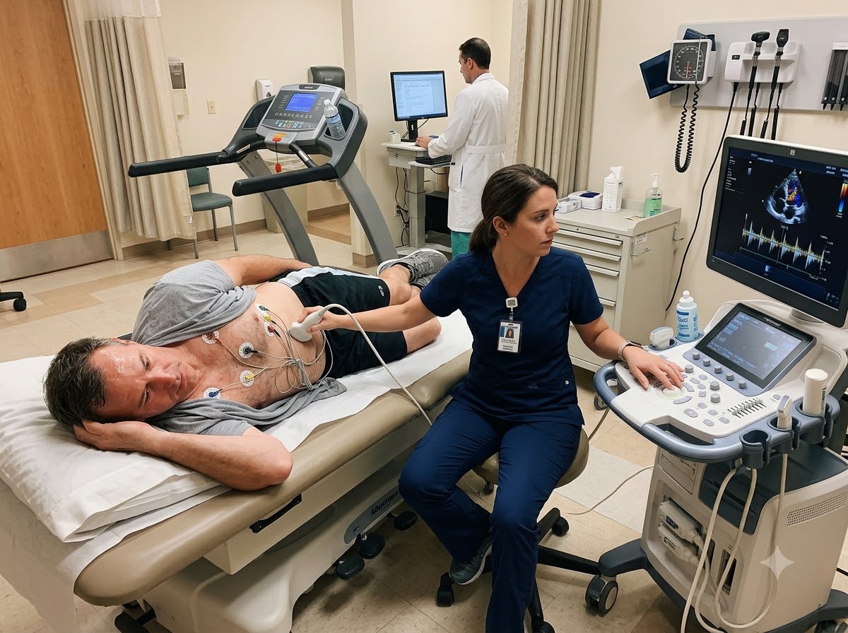
What is a Stress Echo?
A stress Echo (stress echocardiography) joins two tests into one. An Echo is an ultrasound of the heart — the same kind of scan used in pregnancy — that shows the heart walls moving and pumping. A stress test makes the heart beat faster and harder. In a stress Echo, the doctor records Echo pictures before the stress and again immediately after it, and compares the two.
A healthy heart muscle squeezes even more strongly when it works hard. If a heart artery is narrowed, the part of the muscle it supplies cannot get enough blood during stress, and that wall moves weakly or lags behind. Doctors call this a wall motion change. Seeing it directly on the screen is what makes this cardiac stress test so useful.
Think of it like this: a car engine may sound fine while idling in the parking lot. To find a weak part, a mechanic listens to it while driving uphill. A stress Echo watches your heart “driving uphill”.
Two ways to stress the heart — and what a contrast Echo adds
Exercise stress Echo: you walk on a treadmill (or pedal a cycle) while your ECG and BP are watched. As soon as you reach the target heart rate or have to stop, you lie down quickly so the Echo images can be taken within a minute or two, while the heart is still working hard.
Dobutamine stress Echo: if you cannot walk well — because of knee or back pain, weakness, a stroke or lung disease — a medicine called dobutamine is given slowly through a drip in the arm. It makes the heart beat faster and stronger as if you were exercising, while you lie on the couch. Echo pictures are taken at each step.
Contrast Echo: in some people the ultrasound picture is not clear enough — for example in patients who are overweight or have lung disease, where air or fat blocks the view. A contrast agent made of tiny, harmless gas micro-bubbles is then injected into an arm vein. These bubbles light up the inside of the heart on the screen, so the edges of the heart walls are seen much more clearly. Contrast can be used with a stress Echo or with a resting Echo. The bubbles break down in a few minutes and the gas is breathed out.
Who Needs a Stress Echo?
Why is a stress Echo done? Mostly to find out whether chest pain or breathlessness that comes on effort is due to a blockage in the heart arteries, and how serious it is. Your cardiologist decides whether this test is right for you.
Symptoms where a stress Echo can help
Common reasons doctors ask for it
- Stress echo for chest pain when the cause is not clear
- TMT result is unclear, or the resting ECG makes TMT hard to read
- Checking how serious a known blockage is
- Follow-up after angioplasty or bypass surgery if symptoms return
Other situations
- People who cannot walk on a treadmill (dobutamine test)
- Heart fitness check before a major operation
- Some valve problems, to see how the valve behaves on exercise
- Stress echo for breathlessness with an unclear cause
Not for chest pain happening right now. A stress Echo is a planned test. If you have severe chest pain at rest, pain with sweating, or pain spreading to the arm or jaw, call +91 77778 65621 or come to our 24x7 emergency immediately.
What Does Stress Echocardiography Detect?
Because it shows both the heart’s electrical response (ECG) and its actual movement (Echo), a stress Echo gives a fuller picture of how your heart copes with effort.
Narrowed heart arteries
A wall that moves well at rest but weakly after stress points to reduced blood supply (ischemia) from a blockage.
Heart blockageWhich part of the heart is affected
The wall motion pattern helps the doctor judge which artery is likely involved and how large the area at risk is.
Damage from an old heart attack
Areas that do not move at rest or on stress may be scarred from an earlier heart attack.
Heart attack carePumping strength on effort
Shows whether the overall heart function rises normally with exercise or falls, which matters in weak hearts.
Heart failureValve behaviour under load
In some narrowed or leaking valves, the test shows how pressures and flow change during stress.
Valve diseaseYour exercise capacity
How long you could walk, your heart rate and BP response and any symptoms — all useful for planning treatment.
What a stress Echo cannot do: it does not show the arteries themselves, so it cannot measure the exact percentage of a blockage. If the test suggests a significant problem, your doctor may advise a coronary angiography. A normal stress Echo is reassuring, but no test is perfect — tell your doctor if symptoms continue.
What Should I Expect During a Stress Echo?
How long does stress Echo take? Plan for about 45 to 60 minutes at the centre, including getting ready, the stress part and recovery. The exercise itself usually lasts only a few minutes.
Check-up and resting Echo
The team asks about your symptoms and medicines and checks your BP. ECG stickers are placed on your chest and resting Echo images are taken while you lie on your left side.
IV line if needed
For a dobutamine test, or if contrast is planned, a small cannula (drip needle) is placed in an arm vein.
The stress part
You walk on the treadmill, which gets a little faster and steeper every few minutes — or the dobutamine dose is raised step by step while you lie down. Your ECG, BP and symptoms are watched throughout.
Echo images right after stress
With exercise, you move quickly back to the couch so the images are captured while the heart is still beating fast. With dobutamine, images are taken at each stage.
Recovery and rest
Your heart rate and BP are watched until they settle. Most people can go home the same day and return to normal activity, unless the doctor advises otherwise.
Tell the team at once if you feel chest pain, severe breathlessness, dizziness or any discomfort during the test. The stress can be stopped immediately. You may feel your heart pounding with dobutamine — this is expected and passes within minutes after the drip is stopped.
How to Prepare for a Stress Echo — and Is It Safe?
Preparation is simple, but it matters for an accurate result. Our team will tell you the exact instructions when you book.
Good to do
- Fast or have only a light meal, as your doctor advises
- Wear comfortable clothes and walking shoes
- Bring your medicine list and old ECG, Echo and TMT reports
- Ask your doctor which medicines to continue or pause
- Come with a family member if possible
Better to avoid
- Tea, coffee, cola or chocolate for some hours before, if asked
- Smoking on the day of the test
- A heavy meal just before the test
- Stopping any medicine on your own without asking the doctor
- Body oil or lotion on the chest
Is stress Echo safe?
For most people, yes. It uses ultrasound, not X-rays, and it is done under close watch by a trained team with emergency medicines and equipment ready. Common, short-lived effects include breathlessness and tiredness from exercise, or a pounding heartbeat, flushing, tingling or headache with dobutamine.
Like any stress test, there is a small risk of chest pain, a drop or rise in BP, or an irregular heartbeat during the test, and very rarely a heart attack or a serious rhythm problem. The contrast agent can rarely cause an allergic reaction. These events are uncommon, and the team is trained to handle them immediately. Tell us beforehand if you have had any reaction to an injection, or if you are pregnant.
What is the difference between TMT and stress Echo?
Both tests put the heart under stress. The difference is what the doctor looks at while it is working hard.
| TMT (Treadmill Test) | Stress Echo | |
|---|---|---|
| What is checked | ECG and BP changes during exercise | ECG and BP plus ultrasound pictures of the heart walls |
| Shows heart wall movement | No | Yes — before and after stress |
| If you cannot walk | Not possible | Can be done with a medicine (dobutamine) |
| When the resting ECG is abnormal | Result can be hard to read | Echo pictures still give useful information |
| Time needed | About 30–45 minutes | About 45–60 minutes |
| Usually used for | First stress test for chest pain on effort | When more detail is needed, or TMT is unclear |
Which one is right for me? Many patients start with a TMT. Your cardiologist chooses a stress Echo when more detail is needed or a TMT will not give a reliable answer.
What Happens After Your Stress Echo Report?
Your cardiologist looks at the stress Echo together with your symptoms, risk factors and earlier reports. In general, there are three possible paths:
- Normal stress Echo: a significant blockage is unlikely. You get advice on BP, sugar, cholesterol, exercise and a plan for follow-up.
- Small or borderline change: medicines and lifestyle changes may be started, with close follow-up of symptoms.
- Clear signs of reduced blood supply: the doctor may advise a coronary angiography to see the arteries, and angioplasty if needed.
Tip: write down any symptoms you felt during the test — when they started and how they felt. This helps the doctor match your symptoms with the pictures.
Why Get Your Stress Echo at Prathmesh Cardiac Institute?
A stress Echo needs a skilled Echo team and quick decisions during the test. Here it is done in a hospital that treats heart attacks round the clock.
Done and read by heart specialists
DM cardiologists plan your test, watch it and interpret the images.
Exercise or medicine option
If walking is difficult, a dobutamine stress Echo can be planned instead.
Contrast when pictures are unclear
Contrast Echo can be used if your doctor feels clearer images are needed.
Safety first
Continuous ECG and BP watch, with emergency medicines and equipment at hand.
Cath lab under the same roof
If the test shows a serious blockage, angiography and angioplasty are available in the same hospital.
Easy to reach in Raipur
NH-30, Amlidih Road, Deopuri — convenient for patients from Raipur and nearby districts.
Who Reads Your Report
Your report is studied by a DM Cardiology specialist, who explains it to you in plain words and tells you what to do next.
800 × 1000 px · JPG

Dr. Gaurav Tripathi
Performs angiography and angioplasty, and advises on next steps when a stress Echo suggests a blockage.
800 × 1000 px · JPG

Dr. Sulabh Chandrakar
Sees patients with effort chest pain and breathlessness, and explains stress Echo findings in simple words.
Stress Echo — Questions Patients Ask Us
A stress Echo is an ultrasound of the heart taken at rest and again right after the heart is made to work harder, either by exercise or by a medicine. Comparing the two sets of pictures shows whether any part of the heart muscle is short of blood during effort.
It is mainly done to find out whether chest pain or breathlessness on effort is caused by a blockage in the heart arteries. It also helps judge how serious a known blockage is, and how the heart and valves behave under load.
It detects heart walls that move weakly during stress because of reduced blood supply, damage from an old heart attack, the heart’s pumping strength on effort and, in some cases, how a narrowed or leaking valve behaves during exercise.
People with chest pain or breathlessness on effort, those whose TMT was unclear or whose resting ECG makes TMT hard to read, patients who cannot walk on a treadmill, and some patients before major surgery or after angioplasty. Your cardiologist decides if it suits you.
A TMT only looks at ECG and BP changes during exercise. A stress Echo adds ultrasound pictures of the heart walls before and after stress, so it gives more detail and can be done with a medicine if you cannot walk.
Yes, for most people. It uses no radiation and is done under close watch with emergency care ready. There is a small risk of chest pain, BP changes or an irregular heartbeat during the test, and very rarely a heart attack, which the team is trained to handle.
Plan for about 45 to 60 minutes at the centre. The exercise or medicine part usually lasts only a few minutes; the rest of the time is for preparation, the resting and stress images, and recovery.
Resting Echo images are taken first, then you walk on a treadmill or receive dobutamine through a drip while your ECG and BP are watched. Echo images are taken again immediately after stress, and you rest until your heart rate settles.
A contrast Echo uses an injection of tiny, harmless gas micro-bubbles into an arm vein to make the inside of the heart show up brightly on ultrasound. It is used when normal pictures are unclear, for example in overweight patients or those with lung disease.
Usually you are asked to fast or eat only a light meal a few hours before the test, and to avoid tea, coffee and smoking. Ask your doctor about your medicines, especially BP, heart and diabetes medicines, as some may need to be adjusted.