
What Is Coronary Angiography?
Coronary angiography is a test, not an operation. Three main arteries sit on the surface of your heart and supply blood to the heart muscle itself. Angiography is the only test that shows these arteries directly — where they are narrowed, by how much, and how long the narrowed segment is.
A thin, soft tube called a catheter is passed from an artery in your wrist (or sometimes the groin) up to the mouth of the heart arteries. A dye that shows up on X-ray is injected through it, and a camera records moving pictures while the dye flows. On the screen the arteries look like branches of a tree — wherever the dye cannot pass, that is the blockage.
Because it shows the blockage directly rather than guessing from symptoms or from an ECG, angiography is called the gold standard test for coronary artery disease. Everything that comes after — medicines only, angioplasty with a stent, or bypass surgery — is decided from these images.
The whole test is done through a puncture about the size of a ballpoint-pen tip. There is no cut, no stitch, and you are awake and talking throughout. In most cases the arm or leg is not even bandaged beyond a small pressure band.
What does "70% blockage" on the report mean? The percentage is how much of the artery's channel is closed. At 70%, roughly a third of the original channel is left. Below about 50% most people have no symptoms and medicines are enough. Above 70% symptoms usually appear on walking or exertion. But the number alone never decides treatment — where the blockage sits, how many arteries are involved and how well your heart is pumping matter just as much. This is why two people with "70% blockage" can correctly be advised two different treatments.
Why Is Coronary Angiography Done? — Indications
Angiography is advised when a doctor needs certainty. Symptoms, ECG, echo and TMT can all suggest a blockage, but none of them can tell you exactly which artery is blocked and how severely. The test is done to answer very specific questions:
- Is there really a blockage? — A TMT or stress echo can be falsely positive — angiography settles it. Many patients are told after angiography that their arteries are clean, and no stent is needed at all.
- Which artery, and where in it? — A blockage at the start of the main left artery is far more serious than the same blockage in a small side branch, even at the same percentage.
- How many arteries are involved? — Single-vessel disease is usually treated with a stent; three-vessel disease, or disease of the left main artery, may be safer with bypass surgery.
- Is it suitable for a stent? — The images show whether the blockage is short and soft, or long, calcified or fully closed — which decides whether angioplasty is straightforward, complex, or better handled surgically.
- During a heart attack — In an ongoing heart attack, angiography is done immediately and the blocked artery is opened in the same sitting (primary angioplasty). Here it is an emergency, not a planned test.
- Before heart or major surgery — Before valve surgery, and before some major non-cardiac operations in high-risk patients, the heart arteries are checked first so there is no surprise during surgery.
Who May Need Coronary Angiography?
Your cardiologist usually advises angiography after simpler tests point towards a blockage, or when the picture does not add up. Common situations are:
- Chest pain or heaviness on walking that settles with rest — The classic pattern of angina, especially if it is getting worse or coming on with less effort than before.
- A positive TMT or stress echo — The treadmill test or stress echo suggests the heart is short of blood when it works harder.
- After a heart attack — To see the artery that caused it and check the other two.
- Unexplained breathlessness or a weak pumping heart (low EF) — To find out whether blocked arteries are the reason the pumping has dropped.
- Diabetes with suspicious symptoms — People with diabetes often have silent blockages — breathlessness or unusual tiredness may be the only sign, with no chest pain at all.
- Before valve surgery or other heart surgery — Standard practice above a certain age, so blockages can be treated at the same time if needed.
A note on when it is not needed: Angiography is not a routine screening test and is not done just because someone is anxious about the heart. If your symptoms are mild and the non-invasive tests are normal, the right advice is medicines, risk-factor control and follow-up. If a doctor suggests angiography and you are unsure, it is completely reasonable to ask "what will change in my treatment depending on the result?" — a good cardiologist will have a clear answer.
Types / Variations of Coronary Angiography
Radial (wrist) angiography
The route used for most patients today. The puncture is in the wrist artery, you can sit up and eat almost immediately, and bleeding problems are far less common than with the groin. The hand has a second artery supplying it, so blood flow is safe even while the wrist artery is being used.
Femoral (groin) angiography
Used when the wrist arteries are small, too twisty, or already used before, and in some emergencies. It requires lying flat for a few hours afterwards so the puncture site seals properly.
Angiography with FFR or IVUS
When a blockage looks borderline — not clearly mild, not clearly severe — a pressure wire (FFR) or an ultrasound probe (IVUS) is passed into the artery during the same test to decide whether a stent is genuinely needed.
Graft angiography
For patients who have had bypass surgery earlier. The bypass grafts are studied as well as the original arteries, which takes a little longer.
Angiography plus angioplasty in the same sitting
If a significant blockage is found and you have consented in advance, the stent can be placed immediately — no second procedure, no second puncture, no second admission.
CT coronary angiography
A different test altogether: a CT scan with dye given through a vein in the arm. There is no catheter. It is useful mainly to rule out disease in people at lower risk, but it cannot treat a blockage, and heavy calcium can make the images hard to read.
Your cardiologist will tell you which type or technique is right for you after reviewing your reports.
Benefits of Coronary Angiography
- It gives a definite answer — No other test shows the coronary arteries this clearly. You stop guessing and start treating the actual problem.
- It prevents unnecessary treatment — Many patients walk out knowing their arteries are normal, and that their chest pain is not from the heart — which is itself a result worth having.
- Treatment can happen in the same sitting — If a stent is needed, it can usually be placed immediately, saving a second admission and a second puncture.
- It makes the right choice between stent and surgery possible — Only the images can show whether angioplasty or bypass gives you the better long-term result.
- It is quick, and recovery is fast — Around 20–30 minutes, local anaesthesia only, and most people are home the same day or the next morning.
How Coronary Angiography Is Performed
Angiography is done in the cath lab, a clean room like an operating theatre with an X-ray camera that moves around the table. You will be awake throughout and can speak to the team at any time. Here is exactly what happens, step by step.
You are prepared in the day-care room
An IV line is put in your arm, your wrist or groin is cleaned and shaved if needed, and ECG stickers are placed. You change into a gown and remove any jewellery or metal items.
Local anaesthetic is injected
In the cath lab, the area is cleaned again and covered with a sterile sheet. A small injection numbs the skin at the wrist (or groin). This is the only part that stings, and it lasts a few seconds.
A thin sheath is placed in the artery
A short plastic tube (sheath) is inserted into the numbed artery. Everything else passes through this sheath, so the artery is punctured only once. You feel pressure, not pain.
The catheter is guided to the heart
A soft catheter is advanced through the artery up to the mouth of the coronary arteries. There are no nerve endings inside arteries, so you do not feel the catheter moving.
Dye is injected and pictures are taken
Dye is injected in short bursts while the camera records. You may be asked to hold your breath for a few seconds or to cough. Many patients feel a brief warm flush — that is normal and passes in seconds.
Both main systems are studied
The left and the right coronary systems are filmed from several angles, because a blockage can hide when viewed from one direction only. If a pressure wire or ultrasound is needed, it is used now.
The images are reviewed with you
The cardiologist looks at the recorded images and tells you what he can see — often on the screen right there. If a stent is needed and was consented, it is done in the same sitting.
The sheath is removed and pressure applied
The sheath comes out and a pressure band is applied at the wrist, or firm pressure then a dressing at the groin. The band is loosened in stages over a few hours.
You will not be asleep. Many patients expect to be unconscious like in surgery. You are not. You stay awake, you can talk, and most people find it far less frightening than they expected. If you are very anxious, tell the team — a mild calming medicine can be given.
Preparation Before Coronary Angiography
Preparation is simple, but two things matter a lot: your medicine list and your kidney reports. Bring every strip or box of medicine you take, and all previous ECG, echo, TMT and blood reports, including any old angiography CD.
Please do
- Fast for about 4–6 hours before the test — no solid food. Small sips of water are usually allowed; confirm with your doctor.
- Take your blood pressure and heart medicines with a sip of water unless you are told otherwise.
- Tell the team about any allergy, especially a previous reaction to contrast dye, iodine or any drug.
- Tell them if you are, or might be, pregnant.
- Bring your spectacles, your medicine list, previous reports and your insurance or scheme papers.
- Arrange for someone to come with you and to take you home afterwards.
- Wear loose clothes and remove rings, bangles and a watch from the hand that will be used.
Please avoid
- Do not stop any medicine on your own — especially blood thinners. The doctor will tell you which to stop and when.
- If you take metformin for diabetes, ask when to stop and restart it, as it may need to be paused around the dye.
- Do not take your morning diabetes tablet or insulin dose without asking, since you will be fasting.
- Avoid a heavy meal the night before.
- Do not smoke on the day of the test.
- Do not apply oil or cream on the wrist or groin area.
If your kidney reports are abnormal The dye is cleared by the kidneys. If your creatinine is raised, the test can still be done safely in most cases — you will be given extra IV fluids before and after, the smallest possible amount of dye will be used, and certain medicines will be paused. Tell your doctor if you have kidney disease, so this is planned in advance.
Tests Required Before Coronary Angiography
- Blood tests — haemoglobin, creatinine, blood sugar, clotting profile — Haemoglobin and clotting show whether bleeding is likely; creatinine tells the team how your kidneys will handle the dye and how much fluid you need.
- ECG — Records your rhythm on the day and gives a baseline to compare with afterwards.
- 2D Echo — Shows the pumping strength (EF) and the valves, which affects how the procedure is planned and what the images mean.
- TMT or stress echo (usually done earlier) — This is often the test that led to the angiography. Bring the report — it helps match a blockage with the area of heart muscle that is short of blood.
- Infection screening — HIV, hepatitis B and C — Routine before any catheter procedure, as per hospital protocol.
- Chest X-ray — Done if advised, especially if you have breathlessness or lung problems.
If you have had these tests recently somewhere else, bring the reports — many of them do not need to be repeated. That alone can save you both money and a day's work.
Anaesthesia / Sedation Used in Coronary Angiography
Only local anaesthesia is used. A small amount of numbing medicine is injected into the skin over the artery in your wrist or groin. You feel a sting for a few seconds, then the area goes numb.
You stay fully awake. You can talk to the doctor, answer questions and tell the team if anything feels uncomfortable. There is no breathing tube, no ventilator and none of the grogginess that follows general anaesthesia.
If you are very anxious, a mild sedative can be given through the IV line to help you relax. You will still be awake, just calmer. Say so beforehand rather than holding it in — it is a very common request.
What you will actually feel: pressure at the puncture site, a warm flush through the body when the dye goes in, and sometimes a few extra or skipped heartbeats which settle on their own. Pain inside the chest during the test is uncommon; if you feel any, tell the team immediately.
How Long Does Coronary Angiography Take?
The angiography itself usually takes about 20–30 minutes from the first injection to the pressure band. If pressure-wire (FFR) or ultrasound (IVUS) checks are added, allow another 10–20 minutes. If an angioplasty is done in the same sitting, the total time goes up to roughly an hour or more.
The time you spend in the hospital is longer than the procedure itself. Plan for most of the day: admission and paperwork, blood tests if not already done, the procedure, and then a few hours of observation while the pressure band is gradually released.
Hospital Stay / Admission for Coronary Angiography
For a planned angiography through the wrist, most patients are admitted in the morning and go home the same evening, or stay one night and leave the next morning. You will not be moved to an ICU for a routine angiography.
After a wrist procedure you can sit up, eat and walk to the toilet within a short time. After a groin procedure you must lie flat with the leg straight for about 4–6 hours, so a one-night stay is more common.
If an angioplasty is done in the same sitting, the stay becomes about 1–2 days so that you can be monitored and started on the right medicines.
Risks & Possible Complications of Coronary Angiography
Angiography is one of the safest invasive tests in medicine, and serious complications are rare. It is still a procedure on the heart's arteries, so the risks should be understood rather than brushed aside. Most of what happens is minor and settles on its own.
Common and minor
- Bruising, a small lump or tenderness at the puncture site for a few days — common and harmless.
- A warm flush or a metallic taste when the dye is injected, lasting a few seconds.
- A brief feeling of extra or skipped heartbeats during the catheter movement.
- Mild soreness in the wrist or groin for a day or two.
Uncommon or rare
- A larger collection of blood (haematoma) at the puncture site needing pressure and observation.
- An allergic reaction to the dye — itching or rash is uncommon, a severe reaction is rare and the team is equipped for it.
- A temporary drop in kidney function from the dye, mainly in patients who already have kidney disease or diabetes.
- Injury to the artery, a clot, or a rhythm disturbance — rare, and treated immediately in the cath lab.
- Heart attack or stroke during the procedure — very rare, in the range of a fraction of one percent for a routine diagnostic test.
Radiation Angiography uses X-rays, so there is a small radiation exposure — similar in order to other imaging tests, and far smaller than the risk of leaving a serious blockage undiagnosed. Modern cath labs use low-dose settings, and the team keeps screening time to a minimum. For a pregnant patient the test is avoided unless it is an emergency.
Recovery After Coronary Angiography
You rest in the day-care room. The wrist band is loosened in stages, or in the case of a groin puncture you lie flat with the leg straight. You can eat and drink normally once the doctor allows it, and you are encouraged to drink extra water to flush the dye out.
Most people go home the same evening or the next morning. Someone else should drive. You will be given a discharge summary — keep it safely, as it contains the findings and the plan.
Keep the puncture site dry. Avoid lifting anything heavy with that arm, or climbing stairs repeatedly after a groin puncture. Drink plenty of water unless your doctor has restricted fluids.
The dressing can usually be removed and you can bathe normally — ask at discharge for the exact day. A bruise may appear and then fade over a week or two; this is expected.
After a wrist angiography with no stent, most people with desk jobs are back at work in 1–2 days. If your work involves heavy lifting or driving long distances, ask your doctor — it is usually about a week.
Recovery takes a little longer and the blood-thinning medicines become essential. Your doctor will give you a separate, clear instruction sheet for that.
Aftercare & Precautions After Coronary Angiography
- Drink plenty of water for 24–48 hours to help the kidneys clear the dye, unless you have been told to limit fluids.
- Keep the puncture site clean and dry, and watch it for swelling or bleeding for the first 2 days.
- Do not lift heavy weights with the arm used, or do strenuous work, for about 2–3 days.
- Take every medicine exactly as prescribed — and never stop a blood thinner on your own, especially if a stent was placed.
- Restart metformin only when your doctor tells you to, usually after the kidney report is checked.
- Keep the discharge summary, the angiography CD and the report together in one file and carry them to every visit.
- Control the things that caused the blockage in the first place: BP, blood sugar, cholesterol, tobacco and weight. Angiography diagnoses, it does not treat.
- Attend the follow-up visit even if you feel completely fine.
Success / Expected Outcomes of Coronary Angiography
Angiography succeeds in showing the coronary arteries in the overwhelming majority of patients, and the result is available immediately — your cardiologist can usually tell you the findings the same day, often while you are still on the table.
What follows depends on what is found. Normal or mildly narrowed arteries mean no stent and no surgery — treatment is medicines, control of BP, sugar and cholesterol, and a repeat assessment only if symptoms change. A single significant blockage is usually treated with angioplasty and a stent. Disease of all three arteries, or of the left main artery, especially with diabetes, is often safer with bypass surgery, and you will be told that honestly even though it means referring you for an operation.
The honest expectation to have is this: angiography gives you certainty and a plan. It does not by itself make the heart better — that comes from the treatment chosen afterwards and from controlling the risk factors for the rest of your life.
Ask for your images Ask for the angiography CD and keep it. If you ever take a second opinion, a cardiologist anywhere can read those images directly and you will not have to repeat the test.
When to Seek Medical Help After Coronary Angiography
Most people have no problems at all after going home. Contact us immediately — or go to the nearest emergency department — if you notice any of the following:
- Fresh bleeding from the puncture site: press firmly on it with your other hand, keep pressing, and call us
- A rapidly growing lump, severe swelling or increasing pain at the wrist or groin
- The hand or leg on that side becoming cold, pale, numb, blue or very painful
- Chest pain, heaviness or pressure, especially with sweating or breathlessness
- Fever, or redness, warmth or discharge from the puncture site
- Passing much less urine than usual, or swelling of the feet, in the days after the test
- Rash, itching, swelling of the face or difficulty breathing — a possible dye reaction
Coronary Angiography — Questions Patients Ask
The cost depends on whether it is done through the wrist or groin, how much dye and how many catheters are needed, whether FFR or IVUS is added, and your room category. We give you a written estimate before admission rather than a figure after the procedure. Government health schemes and most insurance policies cover angiography, and our team checks your eligibility and completes the paperwork with you. If angioplasty becomes necessary in the same sitting, the cost of stents is explained separately and in advance.
Only if a significant blockage is found and you have agreed to it beforehand. Many patients prefer to give that consent in advance so that a second procedure and a second puncture can be avoided. But it is your decision, and you can also ask for the angiography alone, discuss the findings with your family, and come back for angioplasty later — in a stable patient, a short delay is usually safe. If your arteries are found to be clean, nothing further is done.
We use the wrist (radial) route for most patients. Bleeding complications are less common, you can sit up and walk soon afterwards, and discharge is faster. The groin is used when the wrist arteries are too small or twisty, when they have been used repeatedly before, or in certain emergencies. Both routes give exactly the same quality of images — the difference is in comfort and recovery, not in accuracy.
In most such patients it can be done safely, but it is planned differently. You will be given extra IV fluids before and after the test, the smallest useful amount of dye is used, metformin is paused around the procedure, and your creatinine is rechecked afterwards. Tell your doctor about your kidney reports in advance so this plan is ready. The risk of a temporary drop in kidney function is higher in diabetes with kidney disease, but it is usually mild and recovers.
The test itself takes about 20–30 minutes. After a wrist angiography most patients go home the same evening or the next morning. After a groin angiography you need to lie flat for about 4–6 hours, so an overnight stay is more usual. If a stent is placed in the same sitting, plan for about 1–2 days.
After a plain wrist angiography, people with desk jobs are usually back at work within 1–2 days. Avoid driving on the day of the test — someone else should take you home — and avoid heavy lifting with that arm for 2–3 days. For work involving heavy physical labour, or for long-distance travel, ask your doctor at discharge; it is often about a week. If a stent was placed, the advice is different and will be given separately.
The only painful part is the small injection of local anaesthetic, which stings for a few seconds. After that you feel pressure, not pain. The catheter moving inside the arteries cannot be felt, because arteries have no pain nerves inside them. Most patients say afterwards that it was far easier than they had imagined. A warm flush through the body when the dye is injected is normal and passes within seconds.
Sometimes. CT coronary angiography uses dye through an arm vein and no catheter, and it is useful mainly to rule out disease in people at lower risk. But it cannot measure a blockage as precisely when there is heavy calcium, and most importantly, nothing can be treated during it — if it shows a significant blockage you will still need a conventional angiography. Your cardiologist will tell you which is right for your situation.
The X-ray dose in a routine diagnostic angiography is small and is comparable in order to other imaging tests. Modern cath labs use low-dose settings and the team keeps screening time as short as possible. For almost every patient, the risk of leaving a serious blockage undiagnosed is far greater than the radiation risk. In pregnancy the test is avoided unless it is an emergency.
Usually not. A blockage below about 70% is generally treated with medicines and risk-factor control, because a stent in a mild blockage does not improve survival. Where the narrowing is in between, and it is not clear whether it is limiting blood flow, we can measure it directly with an FFR pressure wire during the angiography and decide objectively. If you are told that every narrowing needs a stent, that is a reasonable moment to ask for a second opinion.
No solid food for about 4–6 hours before the test; small sips of water are usually allowed. Take your blood pressure and heart medicines with a sip of water unless you have been told otherwise. Do not take your diabetes tablet or insulin dose without asking, because you will be fasting. Never stop a blood thinner on your own — your doctor will tell you exactly which medicine to pause and when.
Yes. Age alone is not a reason to avoid it. Many patients in their seventies and eighties undergo angiography safely, often through the wrist, which allows them to sit up and move much sooner. What matters more than age is kidney function, overall frailty and other illnesses — all of which are assessed beforehand and factored into the plan.
Why Choose Prathmesh Cardiac Institute for Coronary Angiography?
Experienced hands, the right equipment and honest advice — the three things that matter most for a safe procedure.
Experienced DM cardiologists
Dr. Gaurav Tripathi and Dr. Sulabh Chandrakar — experienced interventional cardiologists who perform Coronary Angiography regularly.
Advanced, modern Cath Lab
3D angiography with IVUS and FFR guidance — the latest equipment for safe, precise procedures.
24x7 emergency & critical care
A fully equipped CCU and an on-call cath lab team, because heart emergencies do not wait for OPD hours.
Transparent pricing & insurance help
Costs are explained clearly before the procedure, and our team helps you check what your insurance or government health scheme covers.
Complete heart care under one roof
ECG, echo, TMT, angiography, procedures and follow-up in one place — from diagnosis to treatment.
Metro-level care, close to home
The same technology and technique used in metro cardiac centres — without travelling far with a sick patient.
Specialists & Facilities Available
Your procedure is planned and performed by our senior DM Cardiology consultants. You can read their full profiles before your visit.
800 × 1000 px · JPG

Dr. Gaurav Tripathi
Handles complex coronary blockages, CTO, rotablation and emergency angioplasty, with IVUS and FFR guided decision making.
800 × 1000 px · JPG
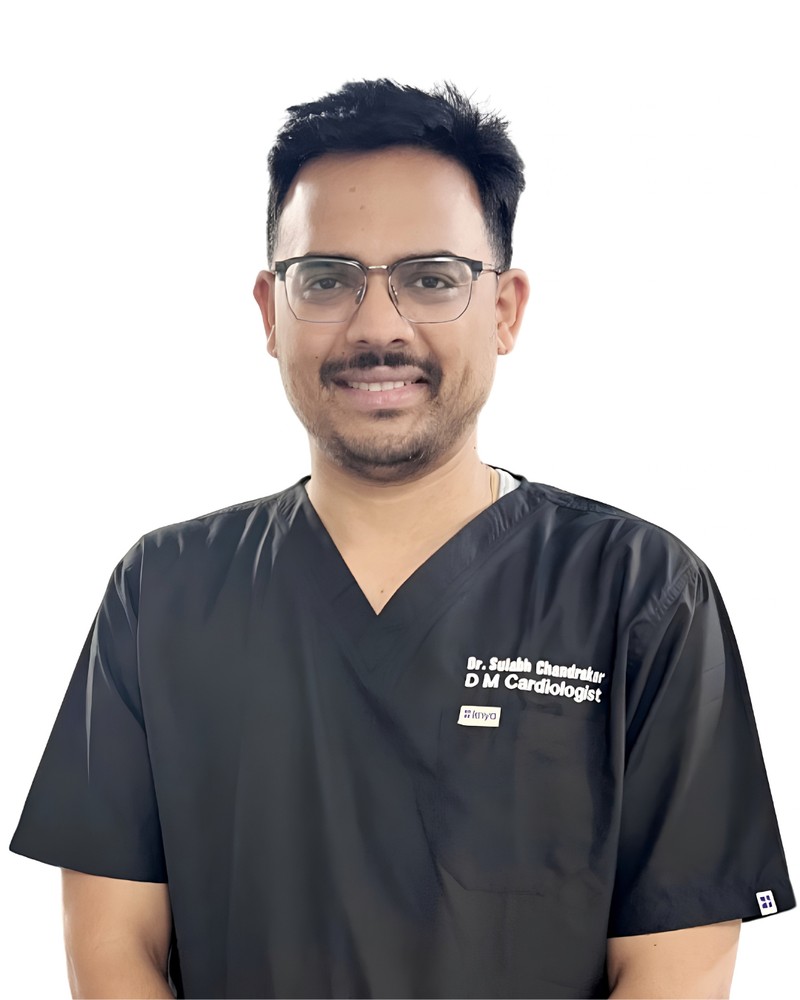
Dr. Sulabh Chandrakar
Performs angioplasty and manages coronary disease with a strong focus on medicines, prevention and long-term follow-up.
Facilities at Prathmesh Cardiac Institute
Book a Consultation / Procedure Appointment for Coronary Angiography
Call or WhatsApp us, or request a call back — our team will confirm a time and tell you what to bring. Bring your previous ECG, echo, angiography reports and current medicine list.