
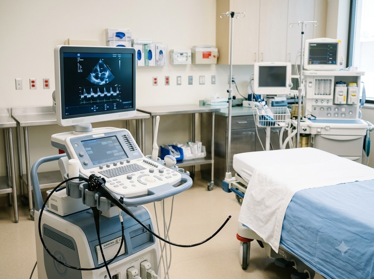
What is a TEE Test?
TEE stands for transesophageal echocardiography. A normal 2D Echo is done from outside, by moving a probe over the chest. In a TEE, a thin, flexible tube with a small ultrasound probe at its tip is gently passed through the mouth into the food pipe (oesophagus). The food pipe runs right behind the heart, so the probe sits very close to it.
Because there are no ribs, lungs or chest fat in the way, the pictures are much sharper — especially of the heart valves, the left atrium (the upper-left chamber) and a small pouch attached to it called the left atrial appendage, where clots like to form. That is why a TEE is often called a detailed heart Echo or advanced heart imaging.
Think of it like this: a normal Echo is like looking at a room through a frosted front window. A TEE is like opening the back door and looking inside from just a step away — the same room, but every corner is clear.
Why is TEE performed when I have already had an Echo?
A 2D Echo answers most questions about the heart. But some details are small or lie at the back of the heart, where the outside view is blurred. Your cardiologist asks for a TEE when an exact answer is needed to decide treatment — for example, whether a clot is present before correcting an irregular heartbeat, or exactly how a valve is damaged before a repair.
When Does a Doctor Recommend TEE?
A TEE is not a routine checkup. It is planned when your doctor needs a closer look at a specific part of the heart.
Situations where a TEE is commonly advised
Most common reasons
- TEE for blood clot evaluation in the left atrium before cardioversion (an electric shock or medicine to restore normal rhythm)
- TEE for heart valve problem — to see exactly how a valve is narrowed or leaking
- Suspected infection on the heart valves (endocarditis)
- Looking for a hole in the heart (ASD or PFO), for example after a stroke
Before or during procedures
- TEE before heart surgery for valve repair or replacement
- Planning a balloon valve procedure (such as BMV)
- Guiding some valve or hole-closure procedures in the cath lab
- Checking an artificial (prosthetic) valve
Not an emergency test for chest pain. If you have sudden chest pain, sudden breathlessness, weakness on one side of the body or difficulty speaking, call +91 77778 65621 or come to our 24x7 emergency at once.
What Does TEE Detect?
A TEE is used to answer specific questions that change treatment. These are the main things it shows clearly.
Clots in the left atrium
Clots in the left atrium and left atrial appendage, which must be ruled out before cardioversion in atrial fibrillation.
Irregular heartbeatValve details
Exactly which part of a valve is narrowed, leaking or torn — key for planning valve repair, replacement or BMV.
Heart valve diseaseInfection on the valves
Small growths (vegetations) and abscesses from endocarditis, which a normal Echo may miss.
Hole in the heart (ASD / PFO)
The size and position of a hole between the upper chambers, and whether it can be closed with a device.
Congenital heart diseaseArtificial valves
Whether a replaced valve is opening and closing well, or has a leak or clot.
The main artery (aorta)
Thickening, plaque or a tear in the wall of the aorta close to the heart.
What a TEE does not do: it does not show blockages inside the heart arteries — that needs a stress Echo, TMT or coronary angiography. It is also not a replacement for a 2D Echo; most patients have a 2D Echo first.
How is a TEE Test Done?
The probe usually stays in for about 15 to 30 minutes. With preparation and recovery, plan to spend a few hours at the hospital.
Check-up and IV line
The team checks your history, allergies and medicines, takes your consent and places a small drip needle in your arm. Dentures are removed.
Throat numbing and sedation
A numbing spray is used at the back of the throat, and a mild sedative is given through the drip so you feel relaxed and drowsy.
The probe is passed gently
You lie on your left side with a mouth guard. The doctor guides the thin probe down; you may be asked to swallow once to help it pass. You can breathe normally throughout.
Pictures are recorded
The probe is turned and moved slightly to view the valves and chambers. Your oxygen, BP and heart rhythm are watched all the time.
Recovery
The probe is removed and you rest until the sedation wears off. You go home with a family member the same day in most cases.
After a TEE: do not eat or drink until the numbness wears off, usually about 1 hour — start with a sip of water. A mild sore throat for a day is common. Because of the sedation, do not drive or operate machinery for the rest of the day.
How Should I Prepare for TEE?
Good preparation keeps the test safe and comfortable. Our team will give you exact instructions at the time of booking.
Please do
- Fast for 6 hours before the test — no food or drinks
- Bring someone to accompany you and take you home
- Ask your doctor about blood thinners, diabetes and BP medicines
- Tell us about any swallowing difficulty, food pipe problem or allergy
- Bring your medicine list and previous Echo reports
Please avoid
- Eating or drinking within 6 hours of the test
- Driving yourself home on the test day
- Eating or drinking before the throat numbness wears off
- Stopping any medicine on your own without asking the doctor
- Wearing jewellery, or leaving dentures in during the test
Is TEE painful?
Most patients do not find it painful. The throat spray and sedation make it comfortable, and many people remember very little of it. You may feel some pressure or a brief gagging feeling as the probe passes, and a mild sore throat afterwards.
TEE is a safe and commonly done test, but it is more than a normal Echo, so it has some rare risks: a reaction to the sedative, a drop in BP or oxygen level, an irregular heartbeat, minor bleeding or injury in the mouth or throat, and very rarely injury to the food pipe. Your doctor will explain these before taking your consent, and the team watches you closely throughout.
What is the difference between TEE and 2D Echo?
Both are ultrasound tests of the heart. The difference is where the probe is placed — and so, how clearly the back of the heart can be seen.
| 2D Echo (TTE) | TEE | |
|---|---|---|
| Where the probe goes | On the outside of the chest | Through the mouth into the food pipe |
| Preparation | None | 6 hours fasting, IV line |
| Sedation | Not needed | Throat spray and mild sedation |
| Picture detail | Good for most questions | Much sharper view of valves and the back of the heart |
| Clots in the left atrial appendage | Usually not seen well | Seen clearly |
| After the test | Go back to normal at once | No food until numbness wears off; no driving that day |
| Usually used as | The first heart scan | A follow-up test for specific questions |
Do I need both? Usually yes — the 2D Echo comes first, and a TEE is added only when your cardiologist needs a closer, more detailed look.
What Happens After Your TEE Report?
The cardiologist who performs the TEE usually tells you the main findings once you are awake, and gives a written report. What happens next depends on why the test was done:
- No clot before cardioversion: the rhythm correction can usually go ahead as planned.
- Clot found: blood thinners are continued and the cardioversion is postponed, with a repeat check later.
- Valve damage, infection or a hole in the heart: the findings guide the choice of medicines, a catheter procedure or surgery.
Tip: keep the TEE report and images with your other heart records. Your surgeon or interventional cardiologist will need them when planning a procedure.
Why Get Your TEE Test at Prathmesh Cardiac Institute?
A TEE needs a skilled operator, careful sedation and close monitoring. Here it is done in a full cardiac hospital with emergency back-up.
Done by heart specialists
DM cardiologists perform your TEE and read the images themselves.
Comfort and safety
Throat numbing, mild sedation and continuous oxygen, BP and ECG monitoring.
Clear explanation
You and your family are told what was found in simple words.
Linked to treatment
Cardioversion, valve procedures and hole-closure planning are handled by the same team.
24x7 cardiac back-up
CCU and cath lab in the same building, round the clock.
Easy to reach in Raipur
NH-30, Amlidih Road, Deopuri — convenient for patients from Raipur and nearby districts.
Who Reads Your Report
Your report is studied by a DM Cardiology specialist, who explains it to you in plain words and tells you what to do next.
800 × 1000 px · JPG
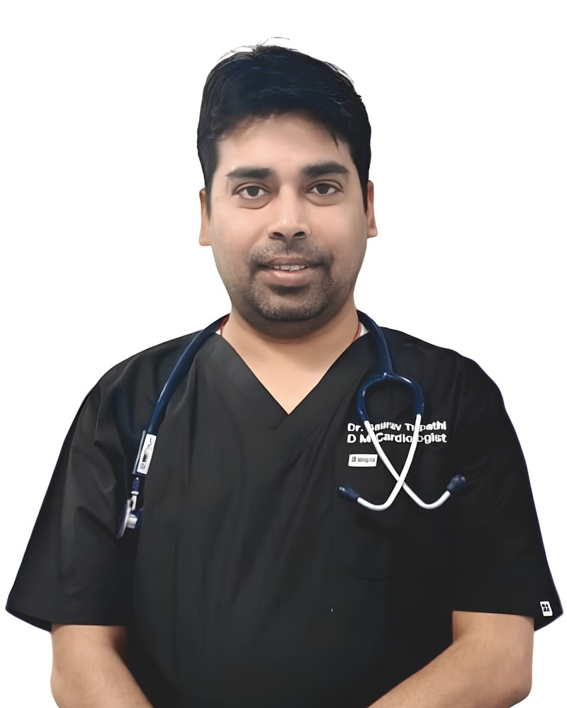
Dr. Gaurav Tripathi
Uses TEE findings to plan catheter-based procedures such as balloon valve treatment and hole closure.
800 × 1000 px · JPG
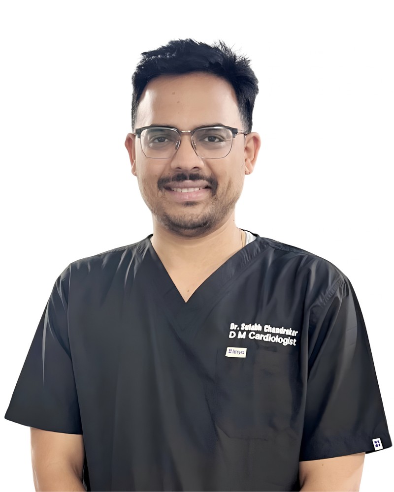
Dr. Sulabh Chandrakar
Sees patients with valve disease and atrial fibrillation, and explains why a TEE is needed and what it showed.
TEE Test — Questions Patients Ask Us
A TEE (transesophageal echocardiography) is an ultrasound of the heart taken from inside the food pipe, which lies just behind the heart. It gives much clearer pictures of the valves and the back of the heart than a normal Echo done over the chest.
TEE is performed when a precise answer is needed that a 2D Echo cannot give clearly — for example to rule out a clot before cardioversion, to check for valve infection, to study a damaged valve before repair, or to look for a hole in the heart.
After fasting, your throat is numbed with a spray and you are given mild sedation through a drip. A thin, flexible probe is gently passed through the mouth into the food pipe and pictures are taken for about 15 to 30 minutes while your oxygen and BP are watched.
TEE detects clots in the left atrium and left atrial appendage, detailed valve damage, infection on the valves (endocarditis), holes in the heart such as ASD or PFO, problems with artificial valves, and disease of the aorta near the heart.
A doctor recommends TEE before cardioversion in atrial fibrillation, when valve infection is suspected, before or during valve procedures, after a stroke to look for a source of clot or a hole in the heart, and when 2D Echo pictures are not clear enough.
Most people do not find it painful. The throat is numbed and mild sedation is given, so you feel relaxed; you may feel brief pressure or gagging as the probe passes and a mild sore throat for a day afterwards.
Do not eat or drink for 6 hours before the test, bring a family member to take you home, and ask your doctor about blood thinners and diabetes medicines. Do not drive for the rest of the day, and do not eat or drink until the throat numbness wears off.
A 2D Echo is done from outside the chest with no preparation. A TEE is done from inside the food pipe with fasting and sedation, and gives far more detailed pictures of the valves, left atrium and back of the heart.
Yes, it is a commonly done and safe test. Rare risks include a reaction to the sedative, a drop in BP or oxygen, an irregular heartbeat, minor throat injury and, very rarely, injury to the food pipe. Your doctor explains these before the test.