
What is Heart Valve Disease?
Your heart has four valves — mitral, aortic, tricuspid and pulmonary. Each one works like a one-way gate. It opens to let blood pass forward, then shuts tightly so blood cannot flow backwards.
Valve disease means one of these gates has stopped working properly. There are only two ways this happens. Either the valve becomes tight and narrow so not enough blood can pass through — doctors call this valve stenosis. Or the valve does not close fully and blood leaks backwards — this is called valve regurgitation. Sometimes both happen in the same valve. Depending on which gate is affected, reports may say mitral valve disease, aortic valve disease, tricuspid or pulmonary valve disease.
Think of the gate of your house. If the gate is half stuck, only one person can squeeze through at a time and a queue builds up outside — that is a narrowed valve. If the gate does not latch and keeps swinging open, whatever you pushed out comes right back in — that is a leaking valve. In both cases the heart has to do extra work every single beat, and over the years that extra work tires the heart muscle out.
Which valve is affected most often in India?
In India, the mitral valve is affected far more often than in Western countries, and usually at a younger age. The reason is rheumatic heart disease — damage left behind years after a childhood throat infection that was never treated properly. Many patients are surprised to hear that a sore throat at age ten caused a valve problem at age thirty-five.
The aortic valve is the other common one, usually affected later in life as calcium slowly deposits on it and stiffens it. This is why aortic valve narrowing is mostly seen after the age of sixty-five.
What Causes Heart Valve Problems?
Valve disease has different causes from blockage. It is not mainly about cholesterol or oily food. These are the real reasons.
You can control these
- Untreated throat infection in childhood — this is the single biggest preventable cause in India. A sore throat with fever must be treated properly, not ignored.
- Stopping rheumatic fever prevention injections halfway — these must be continued for the full duration the doctor advises
- Untreated high blood pressure, which strains the aortic valve over the years
- Untreated infection anywhere in the body that reaches the bloodstream and settles on a valve
- Poor dental hygiene and untreated tooth infections — bacteria from the mouth can reach a heart valve
- Uncontrolled diabetes, which speeds up calcium deposits on valves
You cannot control these
- Increasing age — calcium naturally builds up on valves over decades
- Being born with an abnormal valve, for example a valve with two flaps instead of three (bicuspid aortic valve)
- Past rheumatic fever, once the damage is already done
- Damage after a previous heart attack, which can pull a valve out of shape
- Family history of valve problems
Rheumatic valve disease is largely preventable. If a child in your family gets repeated sore throats with fever, get it treated properly — that one step protects the heart valves for life.
What Are the Symptoms of Heart Valve Disease?
Valve disease is slow. It can sit quietly for years while the heart compensates. Symptoms usually start only once the valve is significantly affected — which is exactly why an early check matters.
Heart valve problem symptoms — early warning signs
- Breathlessness that keeps getting worse over months — first while climbing stairs, then while walking on level ground, and finally even at rest. This gradual worsening is the most typical sign.
- Getting tired far more easily than you used to, without any other explanation
- Feeling your heartbeat fluttering, racing or skipping (palpitations)
- Swelling in the feet, ankles or legs, especially by evening
- Waking up at night short of breath, or needing two or three pillows to sleep comfortably
- Chest discomfort or dizziness while exerting — this is common in aortic valve narrowing
- A doctor hearing a heart murmur (an extra whooshing sound) when listening to your chest, often found during a routine check-up
One point families should know: fainting while exerting, in a person with a known valve problem, is never a small thing. In aortic valve narrowing it is a serious warning sign and needs an urgent cardiology review, not a wait-and-watch approach.
Emergency signs — do not wait, call now
Valve disease itself is usually gradual, but these situations need immediate attention.
- Sudden severe breathlessness, unable to lie flat
- Fainting or near-fainting during activity
- Chest pain along with breathlessness
- Coughing up pink or blood-stained froth
- Very fast or irregular heartbeat with giddiness
- Continuous fever for over a week with a known valve problem
How Is Heart Valve Disease Diagnosed?
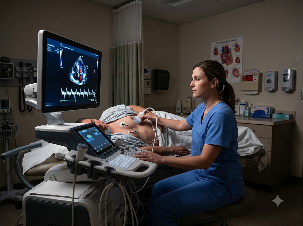
Valve problem diagnosis is mostly done with ultrasound (echocardiography), so it is painless and involves no radiation. Here is what each test involves, so there is nothing to worry about.
Heart Valve Test: 2D Echocardiography
The main test for valve disease. An ultrasound of the heart that shows each valve opening and closing live on screen, and measures exactly how tight or leaky it is.
Read about 2D EchoTEE (Transesophageal Echo)
A closer look at the valve from behind the heart, using a thin probe passed through the food pipe. The throat is numbed first and mild sedation is given, so you stay comfortable.
Read about TEEECG
Records the heart’s electrical activity. It shows whether the extra workload has thickened the heart muscle or upset the rhythm.
Read about ECGTMT (Treadmill Test)
Walking on a treadmill while the ECG runs. It helps judge how much a valve problem is truly limiting you, when symptoms are unclear.
Read about TMTStress & Contrast Echo
An echo taken while the heart is working harder. Useful when the resting echo is borderline and a decision on treatment has to be made.
Read about Stress EchoCoronary Angiography
Done before any valve procedure in older patients, to check whether the heart arteries also have blockages that need treating at the same time.
Read about AngiographyWhen Does Heart Valve Disease Require Treatment?
Not every valve needs replacing — many valve patients never need a procedure at all. The decision depends on how severe the valve problem is, whether it is causing symptoms, and how the heart muscle is holding up. The honest path is step by step.
Watchful follow-up with regular echo — for mild and moderate valve disease
If the valve is only mildly affected and you have no symptoms, no procedure is needed. What you do need is a repeat echo at the interval your doctor advises, usually every one to two years. This is not "doing nothing" — it is catching the right moment before the heart muscle is harmed.
Medicines — to control symptoms and protect the heart
Medicines cannot open a tight valve or seal a leaking one. What they can do is control blood pressure, remove excess fluid so breathing is easier, steady the heart rhythm, and prevent clots. For rheumatic valve disease, long-term preventive penicillin injections stop further damage — these must never be stopped on your own.
Balloon valve procedures (BMV / BAV) — narrowed heart valve treatment without open surgery
For a tight mitral or aortic valve, a balloon can be passed through a small puncture in the leg and inflated inside the valve to open it up. There is no chest cut and no heart-lung machine. Balloon mitral valvotomy in particular gives excellent long-term results in young Indian patients with rheumatic valve disease.
TAVI — valve replacement through a catheter
For severe aortic valve narrowing, especially in elderly patients or those who are high risk for open surgery, a new valve can be placed through a catheter from the leg. The old valve is pushed aside and the new one takes over immediately. Most patients are walking within a day or two.
Open-heart valve surgery — when it is the better choice
For a badly damaged or heavily leaking valve, especially in a younger patient, surgical repair or replacement may give a better lifelong result. In that case we will say so clearly and refer you. Choosing the right treatment matters more than choosing our own procedure.
How the decision is actually made: the cardiologist looks at your echo with you, shows what the valve is doing, and explains why a particular option fits your case. Age, the state of the heart muscle, whether you plan a pregnancy, and whether you can take lifelong blood thinners all change the answer. You should never leave without understanding why one option was chosen over another.
Valve Procedures Available in Raipur
All of these are done in our Cath Lab through a small puncture, without opening the chest.
TAVI / TAVR
A new aortic valve placed through a catheter from the leg. No chest cut, no heart-lung machine. Suited to elderly and high-surgical-risk patients.
Learn moreBMV (Balloon Mitral Valvotomy)
A balloon opens a tight mitral valve. The standard treatment for rheumatic mitral narrowing, with very good long-term results.
Learn moreBAV (Balloon Aortic Valvotomy)
A balloon opens a tight aortic valve. Often used to stabilise a patient before a definitive procedure.
Learn moreBalloon Pulmonary Valvotomy
Opens a narrowed pulmonary valve, most often a problem present since birth and picked up in children or young adults.
Learn moreTEE-Guided Assessment
Detailed imaging from behind the heart before and during a valve procedure, so the device is sized and placed accurately.
Learn moreCoronary Angiography
Routine before valve treatment in older patients, to rule out blockages that should be treated in the same plan.
Learn moreWhy Choose Prathmesh as Your Heart Valve Specialist in Raipur
Valve disease needs accurate measurement and honest timing. Both matter more than anything else.
DM Cardiology consultants
Your echo is read and your procedure performed by the senior consultant himself, not delegated to junior staff.
Advanced Cath Lab with 3D imaging
TAVI, BMV and BAV all done in-house, with the imaging support these procedures demand.
24x7 CCU
Continuous monitoring after any valve procedure, because the first days are when close watching matters most.
Honest timing advice
If your valve does not yet need treatment, we will tell you to wait and re-check. Operating too early helps nobody.
Complete care under one roof
Echo, TEE, angiography, the procedure and lifelong follow-up in one place in Raipur — no running between hospitals.
Rheumatic valve experience
Rheumatic valve disease is common in Central India and needs a different approach from Western textbooks. Our team treats it regularly.
Who Will Treat You
Your case is handled directly by one of our senior cardiologists. You can read their full profile before your visit.
800 × 1000 px · JPG

Dr. Gaurav Tripathi
Performs TAVI, balloon valve procedures and structural heart intervention, with IVUS and FFR guided decision making.
800 × 1000 px · JPG

Dr. Sulabh Chandrakar
Handles valve assessment, medical management, rhythm problems that come with valve disease, and long-term follow-up.
Questions Patients Ask Us Most
Your heart has four valves — mitral, aortic, tricuspid and pulmonary — that work like one-way gates. Heart valve disease means one of them has become tight and narrow (stenosis), or does not close fully and leaks blood backwards (regurgitation). Either way the heart has to work harder with every beat, and over the years this can tire the heart muscle.
The most typical symptom is breathlessness that slowly gets worse over months — first on stairs, then on level ground, and later even at rest. Other signs are getting tired easily, palpitations, swelling of the feet, needing extra pillows to sleep, and chest discomfort or dizziness on exertion. Many people have no symptoms at first, and the problem is picked up when a doctor hears a murmur.
In India the commonest cause is rheumatic heart disease — damage left behind by a childhood throat infection that was not fully treated, which mostly affects the mitral valve. In older people, calcium slowly building up on the aortic valve is the main cause. Other causes include a valve abnormal from birth, infection settling on a valve, and damage after a heart attack.
It often starts when a doctor hears a murmur while listening to the chest. The main test is a 2D Echo, which shows each valve opening and closing and measures how narrow or leaky it is. A TEE gives a closer look when needed, an ECG checks the rhythm, and a TMT or stress echo helps when symptoms and echo findings do not match.
Yes. An Echo is the main test for valve disease — it is the only simple test that actually shows how each valve is working and how severe the problem is. It takes about twenty minutes, causes no pain and involves no radiation. Once valve disease is found, a repeat echo at regular intervals is how your doctor decides the right time for treatment.
Mild and moderate valve disease without symptoms usually needs only regular echo check-ups and medicines where required. A procedure is usually advised when the valve problem becomes severe, when it starts causing breathlessness, chest pain or fainting, or when the echo shows the heart muscle beginning to enlarge or weaken. The aim is to treat before the heart muscle is permanently harmed.
Yes. A mild leak often needs only monitoring with regular echo and medicines to control blood pressure and remove extra fluid. A severe leak that is causing breathlessness, or making the heart enlarge or weaken, is usually treated with surgical repair or replacement of the valve. The right timing is decided from your echo measurements.
In many cases, yes. Mild and moderate valve disease is managed with medicines and regular echo follow-up. A tight mitral or aortic valve can often be opened with a balloon procedure (BMV or BAV) through a small puncture in the leg, and severe aortic narrowing can be treated with TAVI, where a new valve is placed through a catheter without opening the chest. Some badly damaged valves still do better with open surgery, and the cardiologist will explain which option suits you.
A murmur means a doctor heard turbulent blood flow, and many murmurs are completely harmless. The only way to know is an echo, which takes twenty minutes, causes no pain and involves no radiation. If it turns out to be nothing, you stop worrying. If it is something, you have found it early, which is the best possible position to be in.
Not always. After a balloon procedure such as BMV, usually not. After TAVI with a tissue valve, usually only short-term blood thinning plus regular medicines. After a mechanical valve replacement, yes — lifelong, with periodic INR blood tests.
Many women can, but it must be planned. Pregnancy makes the heart pump far more blood, which strains an already affected valve. If you have valve disease and are planning a pregnancy, see a cardiologist before conceiving, because some valve problems are best corrected first and some medicines must be changed.