
How It Started — Slowly, Over Months
There was no dramatic night, no crushing chest pain. It began quietly, almost a year earlier. She would walk to the temple two lanes away and have to stop once on the way, then twice, then she stopped going. Climbing the stairs at home left her holding the railing, breathing hard. The family thought it was age. “She is seventy-eight,” they said. “What else do you expect?”
Then one afternoon, halfway up the stairs, her vision greyed out and she had to sit down on the step. She did not fully faint, but for a few seconds she was not sure where she was. That near-faint was the sign that could not be explained away as weakness.
At another hospital an echo had already shown a valve problem, and the family was told she was too old and too weak for open-heart surgery. They came to us for a second opinion, mainly to ask whether anything at all could be done.
The symptoms she had — and that families wrongly blame on old age
Please note: This is a representative case, put together from the kind of aortic valve case we treat regularly. The details are illustrative and are not the medical record of one identifiable patient. Every heart and every valve is different, and your own treatment can only be decided after your doctor examines you and studies your reports.
What the Tests Showed
Unlike a heart attack, this is not a race against the clock in minutes. Here the job is to measure the valve carefully and plan the procedure properly, so a set of tests is done over a day or two.
ECG — the first basic test
A simple, painless recording of the heart’s electrical activity. It showed signs that the heart muscle had thickened from years of pushing against a tight valve, and no dangerous rhythm problem at rest.
About the ECG test2D Echo — the main test here
An ultrasound of the heart, done with jelly and a probe on the chest. It showed the aortic valve opening was severely narrowed and the pressure the heart had to generate across it was very high. This one test is what tells us the stenosis is severe.
About 2D EchoTEE — a closer look at the valve
A smaller probe is passed into the food pipe, which lies just behind the heart, under sedation. From there the valve is seen far more clearly — its exact shape, how calcified it is, and whether it leaks as well.
About TEECT scan — to size the new valve
A special CT measures the valve opening and the arteries the valve will travel through, down to the millimetre. This is how we choose the right size of valve and the safest route for it. Without this scan, TAVI cannot be planned.
One more test before the decision: a coronary angiography was done to check whether her heart arteries were also blocked, because a tight valve and blocked arteries often go together. In her case the arteries were adequate, so the valve was the only problem that needed fixing. In plain words, severe aortic stenosis means the small door through which blood leaves the heart has stiffened and narrowed, so the heart has to strain at every beat to push blood out through it.
Why TAVI and Not Open-Heart Surgery
A narrowed aortic valve cannot be cured by tablets. No medicine can widen a stiff, calcified valve. Medicines can ease the breathlessness for a while, but the narrowing stays, and severe aortic stenosis with symptoms like hers does not stay quiet for long. The valve has to be replaced.
Traditionally that meant open-heart surgery: the chest is opened, the heart is stopped, a heart-lung machine takes over the circulation, and the old valve is cut out and stitched over with a new one. It is an excellent, long-proven operation — in the right patient. At seventy-eight, frail, with little muscle strength and months of poor eating behind her, she was not that patient. The surgical risk in her case was high, and the hospital she went to first was not wrong to say so.
TAVI (also called TAVR) is the other way. The new valve is folded onto the tip of a thin tube and passed up from the artery in the groin to the heart. It is then opened inside the old narrowed valve, which gets pushed aside and stays there, and the new valve starts working immediately. The chest is never opened. The heart keeps beating throughout. No heart-lung machine is used. That is why it can be offered to people for whom surgery would be too much.
But TAVI is not the right answer for everyone. It depends on the valve’s shape, how calcium is spread on it, the size of the arteries in the legs, the age and general condition of the patient, and what else is wrong with the heart. That decision is not taken by one person. A heart team — cardiologist, imaging doctor and cardiac surgeon together — looks at the echo, the TEE and the CT and decides whether surgery, TAVI, or sometimes only medicines is the honest best option. In her case the team’s answer was TAVI, and the family was told clearly why.
What made TAVI the right choice for her
- Severe narrowing with clear symptoms — breathlessness and a near-faint
- Age and frailty made open-heart surgery high risk
- The CT showed valve and leg artery measurements suitable for TAVI
- Heart arteries were adequate, so only the valve needed treating
- Recovery without a chest wound mattered a great deal at her age
When TAVI is not the answer
- A younger, fit patient — surgery is often still the better long-term choice
- Valve anatomy or calcium pattern unsuitable on the CT scan
- Leg arteries too narrow or twisted for the valve to pass through safely
- Several other problems needing surgery anyway, such as blockages needing bypass
- An active infection in the body or on the valve — that must be treated first
The Procedure, Step by Step
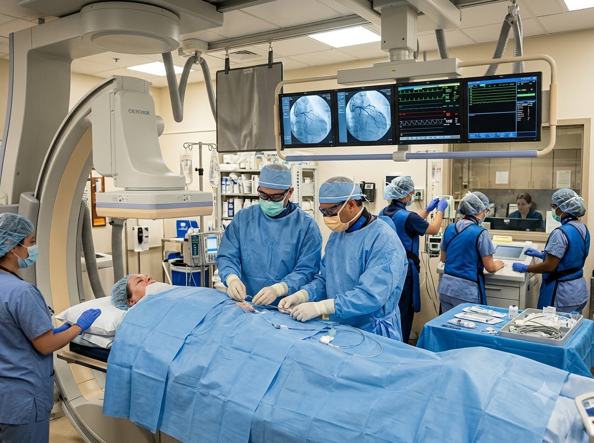
Families picture something terrifying when they hear “valve replacement”. What actually happens in the cath lab is much quieter than that. Here is how her day went.
Preparation and the heart team ready
She came in the previous day. Blood tests, a dental and infection check, and an explanation of the whole plan to her and her sons. Consent was taken only after their questions were answered. In the cath lab she was connected to monitors, with the cardiologist, anaesthetist and the full team present.
The groin is numbed
A local injection numbs the skin at the top of the thigh, where the big artery runs. For TAVI, mild sedation is usually given as well, so the patient is drowsy and comfortable but not put on a ventilator in most cases. There is no cut on the chest at any point.
A thin tube travels up to the heart
A fine tube is passed from the groin artery up to the heart. Arteries have no pain nerves inside, so this part is not painful. The route is followed on X-ray screens the whole way, and her blood pressure and rhythm were watched beat by beat.
The new valve is positioned
The folded valve is brought exactly to the level of the old narrowed valve. Getting this position right, to the millimetre, is the most important moment of the procedure, and it is checked from more than one angle before anything is released.
The valve is opened
The new valve is expanded inside the old one. The stiff old valve is pushed aside against the wall and stays there. The new valve’s leaflets begin opening and closing with her own heartbeat straight away.
Checked, and the tube removed
Pressure across the valve was measured again and an echo was done on the table to confirm the valve was sitting well and not leaking. The tube was removed and the groin was sealed. No stitches on the chest, only a small dressing on the thigh.
What she noticed afterwards: she said the part she had dreaded most — being “operated on” — never happened. She remembered lying on a narrow table, people talking calmly, and a pressing feeling at the top of the leg. By evening she was propped up in bed with her family beside her. The breathlessness she had lived with for months was already noticeably less when she sat up to drink water.
Recovery, Day by Day
This is what families ask us first and what most pages never answer honestly. Recovery after TAVI is quick compared to open surgery, mainly because there is no big wound to heal. But quick does not mean unmonitored.
Same day — sitting up and eating
She was shifted to the CCU for close monitoring of rhythm and blood pressure. She was allowed to sit up and take light food the same evening. The groin site was checked repeatedly for any bleeding or swelling.
Day 1 to 2 — walking in the ward
Out of bed with support, then walking in the corridor. Heart rhythm was watched carefully, because the new valve sits very close to the heart’s own electrical wiring and the rhythm can change in the first days.
Day 3 to 4 — going home
A repeat echo to confirm the valve was working well, a final look at the groin, and the medicine list explained one tablet at a time to her and her daughter-in-law, in writing. Most patients go home after a few days.
Week 1 — moving about at home
Short walks indoors from the first days, gradually out to the veranda and the gate. No lifting, no straining, nothing heavy on the leg that was used. She was walking comfortably around the house within about a week.
Week 4 to 6 — back to her own routine
Follow-up visit with an ECG and echo to see how the valve and the heart muscle are settling. By this point she was going out, climbing the stairs slowly but without stopping, and eating properly again.
Being honest about the risks. TAVI avoids the chest being opened, but it is still a heart procedure and it carries real risks. The one we watch for most closely is a rhythm disturbance: the new valve sits right next to the heart’s natural electrical pathway, and in some patients the beat slows enough afterwards that a pacemaker is needed. There can also be bleeding or a swelling at the groin where the tube went in, and, as with any procedure on the heart and large arteries, a risk of stroke. Kidney function and the new valve’s seal are also checked. These things are discussed with the family before consent, not after — and we do not put a number on them for you here, because the real risk depends on your own heart, your reports and your general health.
The follow-up matters as much as the procedure. A valve is a moving part and it is checked with an echo at planned intervals for the rest of a patient’s life. Medicines after TAVI are usually simpler than after a surgical valve, but they are decided case by case — never copied from another patient and never stopped on your own. Any fever, new breathlessness, swelling of the legs, fainting or a very slow pulse means a call to your cardiologist the same day, not at the next appointment.
What This Means for You
If someone has told you that your mother or father is “too old for heart surgery”, read this part carefully. That sentence is often true about open surgery. It is not the same as saying nothing can be done.
A narrowed valve is a mechanical problem, not a mysterious one. A door inside the heart has gone stiff. Mechanical problems can often be fixed mechanically, and today that can sometimes be done without opening the chest at all. The question to ask is not “is she strong enough for surgery?” but “has a heart team looked at her echo and said what her options are?”
The second thing we want you to take away is about breathlessness in an older person:
- Getting breathless on short walks is not normal ageing — it is a symptom, and it has a cause
- Any fainting or near-fainting on effort in an elderly person needs a heart check, not rest and a tonic
- An echo is the simple first step — painless, no needle, no radiation, and it shows the valves clearly
- If you have been told the valve is severely narrowed, ask specifically about TAVI and about a heart team opinion
- Carry the old echo report and film to the appointment — comparing with an earlier echo tells us a great deal
- Do not wait for the breathlessness to become constant before acting — symptoms mean the valve is already severe
Do not wait if this is happening now. Breathlessness at rest, chest pain, or fainting in an elderly person with a known valve problem needs to be seen today, not next week. Our cardiac emergency is open 24x7 — +91 77778 65621.
Why This Was Possible at Prathmesh
A TAVI is not one doctor’s skill alone. It needs careful imaging, honest decision making and a team that can handle the heart from inside a catheter.
DM Cardiology specialists
The consultant who studies your reports and decides the plan is the one who performs the procedure and sees you in follow-up.
Full valve imaging in-house
ECG, 2D Echo and TEE are done here, so the valve is measured and reviewed carefully before anything is planned.
Cath lab and CCU in one building
Monitoring after a valve procedure continues upstairs in the same hospital — no shifting an elderly patient between hospitals.
Heart team decision, honestly given
If surgery or medicines are the better option for you, we say so. Not every narrowed valve should be treated with TAVI.
Families told what to expect
The procedure, the recovery and the risks, including the chance of needing a pacemaker, are explained in plain language before consent.
Easy to reach from Durg and Bhilai
NH-30, Amlidih Road, Deopuri — a straightforward drive from Durg, Bhilai and the districts around Raipur.
Who Will Treat You
Your case is handled directly by one of our senior cardiologists, who explains every step before it happens.
800 × 1000 px · JPG
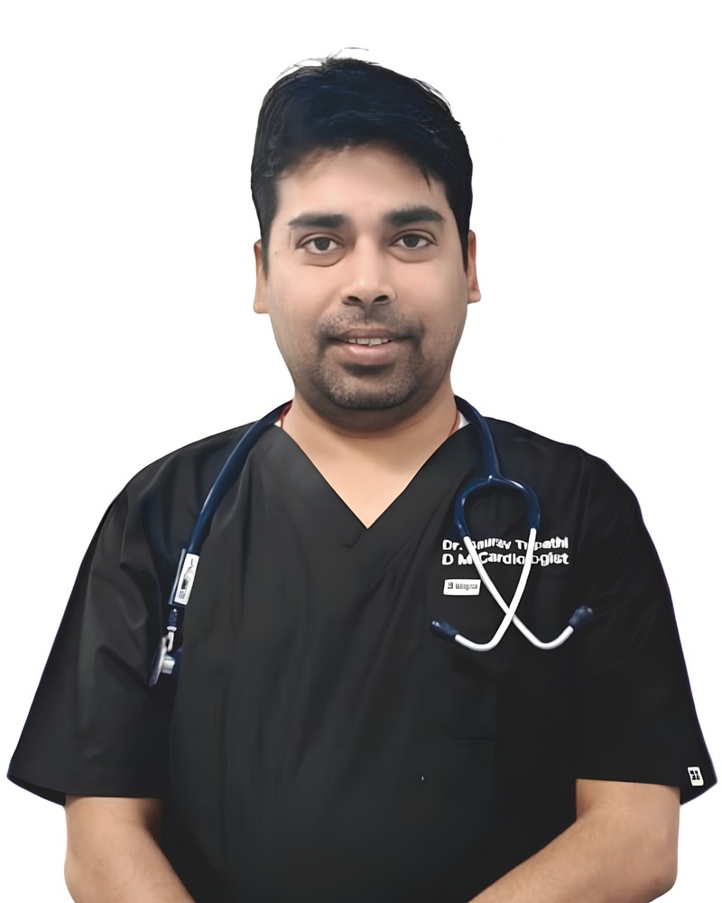
Dr. Gaurav Tripathi
Performs transcatheter valve procedures including TAVI and balloon valvotomy, and plans each case from the echo, TEE and CT measurements.
800 × 1000 px · JPG
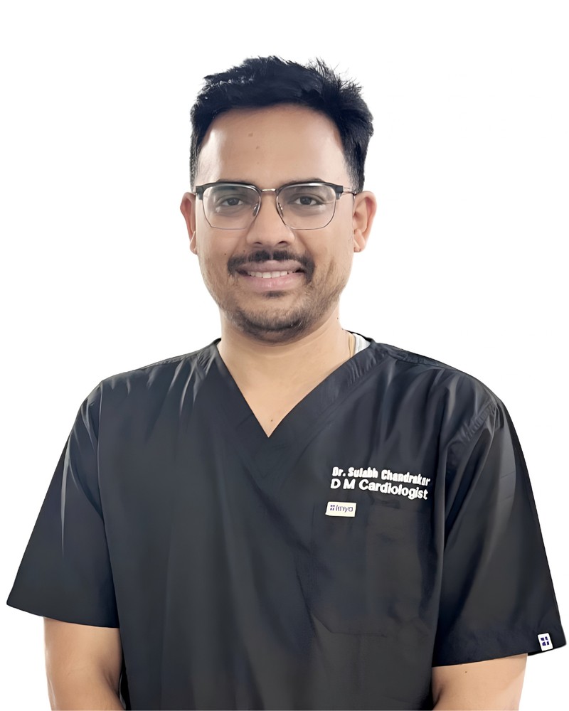
Dr. Sulabh Chandrakar
Assesses valve disease and heart failure, manages rhythm problems after valve procedures, and handles long-term valve follow-up.
Questions Patients Ask Us About This
The aortic valve is the door through which blood leaves the heart to the rest of the body. In aortic stenosis that door becomes stiff and narrow, usually from calcium deposits over many years. The heart then has to push much harder at every beat, which is why patients feel breathless, tired and sometimes faint on effort.
No. The new valve is passed up on a thin tube from the artery in the groin and opened inside the old valve. There is no cut on the chest, no heart-lung machine, and the heart keeps beating throughout. Only a small puncture site at the top of the thigh needs to heal.
Age alone does not decide this. Many patients considered too high risk for open-heart surgery are exactly the people TAVI was developed for. What decides it is the valve anatomy on the CT and echo, the condition of the leg arteries and the overall health of the patient, assessed by a heart team.
No medicine can widen a stiff, calcified valve. Tablets can reduce the breathlessness and control blood pressure and fluid, which helps symptoms for a time, but the narrowing itself remains. In severe aortic stenosis with symptoms, replacing the valve is the treatment that changes the outcome.
As with any heart procedure there are real risks. The main ones are a rhythm disturbance that can sometimes require a pacemaker afterwards, bleeding or swelling at the groin where the tube is inserted, and a risk of stroke. There can also be a small leak around the new valve or an effect on kidney function. Your own level of risk is estimated from your reports and explained to you before consent.
The groin is numbed with a local injection and mild sedation is usually given, so most patients are drowsy and comfortable rather than in pain. Arteries have no pain nerves inside them, so the tube travelling up to the heart is not felt. Patients commonly describe a pressing sensation at the top of the leg and nothing more.
A stay of a few days is common, starting with close monitoring on the first day. Most patients sit up and eat the same day or the next, walk in the ward within a day or two, and are moving comfortably around the house in about a week. There is no large surgical wound to heal, which is the main reason recovery is faster.
The aortic valve sits very close to the heart’s own electrical wiring. When the new valve is expanded, that wiring can get pressed and the heartbeat may become too slow in some patients. If the slow rhythm does not recover, a permanent pacemaker is implanted to keep the heart rate steady. This is why rhythm is monitored closely in the first days.
TAVI valves are made from tissue and, like surgical tissue valves, they do not last forever. They are followed with a planned echo schedule to see how the valve is working over the years. How long any one valve lasts depends on the patient, so your cardiologist will explain what is reasonable to expect in your own case rather than quote a fixed number.
Often yes, but the blockages are assessed first with a coronary angiography. Depending on how severe and where they are, an important blockage may be treated with a stent before or along with the valve procedure, or the heart team may advise surgery for both problems together. This is exactly the kind of decision the team is there to make.